

Ultrasound
Description
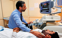 An ultrasound exam is a safe diagnostic procedure that uses very-high-frequency
sound waves to produce an image of many of the internal structures of the body.
These sound waves are harmless and may be used with complete safety, even on pregnant
women, where X-rays would be inappropriate.
An ultrasound exam is a safe diagnostic procedure that uses very-high-frequency
sound waves to produce an image of many of the internal structures of the body.
These sound waves are harmless and may be used with complete safety, even on pregnant
women, where X-rays would be inappropriate.
When high-frequency sound waves pass through soft tissues, echoes are produced at
interfaces, such as the renal capsule or the bile duct wall. Knowing the speed of
sound in soft tissues, the depth of the source of the echo can be calculated from
the time lapse between production of the sound and detection of the echo. This one
-dimensional information is the basis of M-mode ultrasound, which is useful for
studying movement of the heart valves. When an ultrasound beam is scanned through
an arc, combining the direction of the beam with the depth of the echo allows a
two-dimensional image to be produced (B-mode). Modern ultrasound machines permit
rapid scanning, resulting in a 'real-time' image. The ultrasound beam cannot penetrate
bone or air. This restricts the use of ultrasound to soft tissues.
Since fluid provides an ultrasound 'window', the fetus is well seen in the amniotic sac. Ultrasound is the imaging modality of choice in the field of obstetrics. It is the initial modality used in the assessment of patients with hepatobiliary, pancreatic, and renal or gynaecological symptoms. It is valuable in the detection of fluid collections and abscesses, and can be employed to guide aspiration or drainage procedures.
Doppler
When sound waves are echoed from a moving object, e.g. flowing blood cells, the frequency alters. The frequency shift depends on both the velocity and on the direction of movement in relation to the beam. A focal stenosis in an artery produces a high velocity jet, whereas an occluded vessel produces no Doppler signal.
Doppler is increasingly used to evaluate patients with arterial problems, particularly those with lower limb or carotid disease. Doppler imaging can also be used in the evaluation of the venous system.
Procedure
These are the commonly performed ultrasound examination at SMC:
- Abdominal Ultrasound
- Obstetrical Fetal Ultrasound
- Pelvic Ultrasound
- Prostate Ultrasound
- Vaginal Ultrasound
- Small Parts Ultrasound
- Thyroid Ultrasound
- Kidney Transplant Ultrasound
- Vascular Ultrasound
- Ultrasound Guided Procedure
-
Head Scan Ultrasound
Precautions
There is a simple preparation for the following exams:
- Abdominal Ultrasound: Fasting from food 6 hours before the exam (including smoking), but may drink clear water only.
- Obstetrical Fetal Ultrasound: Keeping a full bladder for pregnancies less than 14 weeks.
- Pelvic Ultrasound: Keeping a full bladder
- Prostate Ultrasound: Keeping a full bladder
- Renal (Kidneys & Bladder): Keeping a full bladder
No preparation is necessary for the following ultrasound exams:
- Breast
- Fetal (after fourteen weeks of pregnancy)
- Scrotal (Testicular)
- Thyroid (Neck)
- Doppler (Blood vessels)
-
Head
Location
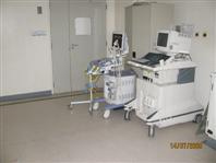
There are 5 high end ultrasound units in five rooms and one unit in in ward 12. Also there are 4 middle range mobile units one of them is portable unit and they are located at the Radiology Department - SMC New Building in Ground Floor.
Dir. Tel. No.: 17284007
Ext. No. : 4007
Equipment
These rooms are equipped with real time linear/sector scanners for general, abdominal, pelvic, obstetric and small parts procedures. In addition to U/S guided biopsies and endovaginal ultrasonography.
Room 12: GE Logiq 9
Room 14: ATL-5000
Room 15: Siemens ACUSON S2000 Breast Volume Scanner;
Room 16: Philips IU 22
Room 18: GE Logiq E9
Ward 11: Philips IU 22
Available Staff
Three Consultant Radiologists cover the daily routine lists and one Chief/Senior Resident covers an emergency list.
Head of Ultrasound Unit Mrs. Wafa Mohd
One Radiologic Technologist: Mrs. Yusra
Two MSA: Mrs.Rabab & Mrs. Fatema
Timing
Sundays to Thursdays from 7:00 AM to 2:00 PM
+24 hours Radiologists on-call